The 45‑Minute Catheter Procedure That Closes Varicose Veins Without Cutting, Stitches, or a Hospital Stay
A Muskoka surgeon explains why sore, bulging leg veins keep getting worse no matter what you try. And the walk‑in, walk‑out treatment most people have never been offered. If your legs feel heavy, swollen, or restless by evening, read this now.
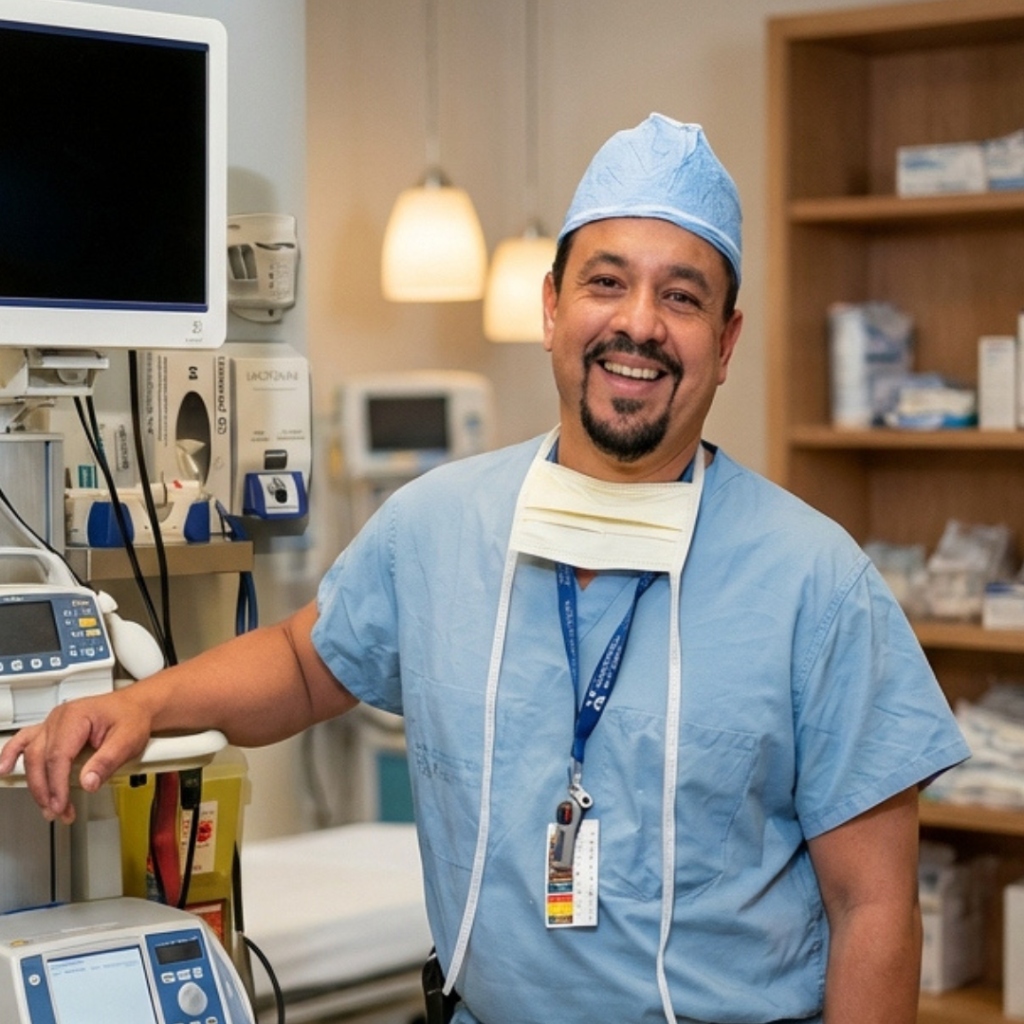

If Your Legs Ache, Swell, or Cramp at Night, This Is for You
You know the feeling. By late afternoon, your legs feel heavy, like they're carrying sandbags. You kick your shoes off and there's a ring pressed into your ankle from your sock. At night, a cramp grabs your calf and jolts you awake. In summer, you quietly stopped wearing shorts.
Maybe you can see them: ropey, bluish veins bulging behind your knee or along your calf. Or maybe you can't see much at all, just the heaviness, the itching, the restless legs that won't settle when you finally sit down.
If any of that sounds familiar, you're not alone. About one in three adults has vein disease. It gets more common with age, pregnancy, jobs on your feet, and family history.
Here's what I want you to know before you read another word: those veins are not just a cosmetic issue, and they will not fix themselves. But there is a fix. Doctors can now close the bad vein from the inside, in under an hour. No cutting. No going under. Most patients walk out the same day. It's backed by over fifteen years of published research.1,2
Most patients I meet have never heard of it. That's why I'm writing this.
Who I Am, and Why I'm Telling You This

My name is Dr. Hector Roldan. I'm a general surgeon, a Fellow of the Royal College of Surgeons of Canada and of the American College of Surgeons, and I've practised in Muskoka since 2007. Vein care has been part of my practice for years. I assess and treat varicose and spider veins right here in the region.
To bring the full range of modern vein treatment to my patients, I've partnered with Dr. Luis Figueroa, a board‑certified vascular surgeon with deep experience in radiofrequency ablation, the catheter procedure this article is about. I'll introduce him properly below. My role here is the one I take with every patient: to explain what's actually going on in your legs, lay out your options honestly, and help you get to the right treatment. That's it.
And after nearly two decades of consultations, I kept seeing the same pattern, and it bothered me.
The Pattern I Couldn't Ignore
A patient sits down in my office. Usually in their 50s or 60s, though sometimes far younger. They've had aching, swollen legs for five, ten, fifteen years. And almost every time, the story is the same:
They were told to wear compression stockings. They were told to elevate their legs. Some were told, politely, that it was "just cosmetic" and to live with it. A few were offered the old surgery, vein stripping, heard the words "general anesthetic" and "weeks of recovery," and understandably walked away.
So they managed. For years. While their legs slowly got worse.
Here's the part that bothered me: a minimally invasive fix for the root cause has existed for over two decades, has been studied in thousands of patients in peer‑reviewed journals, and most of these patients had simply never been offered it. These patients deserved better information. So let me give it to you now.
What Varicose Veins Actually Are (It's Not What You Think)
Most people think of varicose veins as a surface problem: ugly veins that appeared on the skin. That's backwards.
Your leg veins carry blood back up to your heart. That means blood has to travel against gravity. Tiny one‑way valves inside the veins make this work. Each time your calf muscle squeezes, blood moves up. Then the valves snap shut so it can't fall back down.

This is why so many treatments disappoint. If you treat the surface veins but leave the refluxing vein underneath, the pressure is still there. New varicose veins form. The aching comes back. It was never a skin problem. It's a plumbing problem.
Why Everything You've Tried Hasn't Worked (And It's Not Your Fault)
Compression Stockings
Stockings squeeze the leg from outside to help push blood up. They can ease symptoms during the day. But the moment you take them off, the broken valve is still broken. Stockings manage the problem. They cannot fix it. And let's be honest: they're hard to wear every day, especially in summer.
Elevation, Exercise, and "Just Live With It"
All good advice. None of it fixes the broken valve. Gravity wins every time you stand up. And vein disease doesn't stand still. Left alone, the pressure damages more valves. Over years, it can damage the skin itself.
Surface Sclerotherapy Alone
Injecting visible spider or surface veins can work well, and it's a treatment I offer. But when there's untreated reflux in the main saphenous trunk underneath, treating only the surface veins is like mopping the floor with the tap still running. The source keeps feeding new veins.
Traditional Vein Stripping
For decades, the definitive fix was surgery: general anesthesia, incisions at the groin and lower leg, and physically pulling the saphenous vein out of the leg. It works, but at a cost. In randomized trial data, patients who had stripping reported more post‑operative pain and slower return to normal activity than patients treated with the catheter‑based alternative,2 and a 2024 systematic review pooling published studies reported a five‑year recurrence rate of 34.6% after surgical stripping.3 One recognized reason: the trauma of surgery can trigger the growth of new, disorganized veins in the surgical bed, a process called neovascularization.
The Procedure: Radiofrequency Ablation (RFA)
The answer is radiofrequency ablation, sometimes called RFA or catheter‑based vein closure. It has been in clinical use since 1999, refined into its modern segmental form in 2007, and studied in prospective, multicentre, peer‑reviewed trials across Europe and North America.1,2,4
The concept: instead of pulling the diseased vein out of your leg, a thin catheter delivers controlled radiofrequency heat to the vein wall from the inside. The heat causes the collagen in the wall to contract. The vein shrinks, collapses, and seals shut. Over the following months, your body converts it to fibrous tissue and absorbs it. Blood instantly reroutes through the many healthy veins nearby, which is what makes closing the diseased vein safe: your circulation doesn't depend on it. It was working against you.
What Actually Happens, Step by Step
Ultrasound mapping
The physician uses duplex ultrasound to map your veins and confirm exactly where the reflux is coming from. No guesswork; the failed segment is identified before anything else happens.
A single needle entry
The skin is numbed with local anesthetic and the catheter enters the vein through a tiny puncture, usually near the knee or ankle. No incision. No stitches. You are awake and comfortable.
Protective numbing around the vein
A dilute local anesthetic solution (tumescent anesthesia) is placed around the vein under ultrasound guidance. It numbs the area and insulates the surrounding tissue from the heat.
The vein is sealed in segments
The catheter heats the vein wall in short, precisely controlled segments, about 20 seconds each, as it's withdrawn along the vein. The wall contracts and seals behind it. Total treatment time is typically 45 to 60 minutes door to door.
You walk out
A compression stocking goes on, and you're encouraged to walk immediately. Most patients return to normal daily activities within a day or two; strenuous exercise waits about a week.2

The Clinical Evidence
This is not a new or experimental idea. It has been evaluated in prospective trials, randomized comparisons, and long‑term follow‑up studies published in peer‑reviewed surgical journals.
In the EVOLVeS randomized trial comparing radiofrequency closure directly against ligation and stripping, patients in the radiofrequency group reported less post‑operative pain, returned to normal activities faster, and returned to work sooner than the surgical group, with quality‑of‑life scores favouring the catheter procedure through follow‑up.2 A separate multicentre randomized trial (RECOVERY) comparing radiofrequency ablation with endovenous laser found significantly lower pain, bruising, and tenderness scores with radiofrequency at 48 hours, one week, and two weeks.4
| Radiofrequency Ablation | Vein Stripping Surgery | Compression Only | |
|---|---|---|---|
| Anesthetic | Local only | Usually general or spinal | None |
| Incisions | None; single needle entry | Groin and leg incisions | None |
| Setting | Clinic, walk in and out | Operating room | At home |
| Back to daily activity | Typically 1–2 days2 | Days to weeks2 | — |
| Addresses the failed valve | Yes, vein sealed at source | Yes, vein removed | No, symptoms only |
| 5‑year recurrence (pooled data) | 18.7%3 | 34.6%3 | Disease progresses |
Honest expectations matter, so here is the balance: no vein treatment has a zero recurrence rate. Roughly 8% of treated veins in the five‑year European data showed some reopening, and new vein disease can develop elsewhere in the leg over time.1 Minor side effects such as temporary bruising, tightness, or numbness along the treated vein occur in a minority of patients and typically resolve. Serious complications are uncommon in the published literature.1,3 A proper ultrasound assessment determines whether you're a candidate at all.
What Happens If You Keep Waiting
Chronic venous disease doesn't stay still. Physicians grade it in stages, and the honest picture looks like this:
Spider veins, mild aching
Fine surface veins, occasional tiredness in the legs. Easy to dismiss. Often dismissed for years.
Bulging veins, swelling, heaviness, night cramps
Reflux is established. Pressure is remodelling the veins and pushing fluid into the tissue. Symptoms are now daily. This is the window where treatment is simplest and the published outcome data most favourable.
Skin damage and venous ulcers
Years of pressure discolour and harden the skin around the ankle. Eventually the skin can break down into a venous ulcer, an open wound that is slow to heal and prone to returning. As someone who also runs a wound care practice, I can tell you these are far harder to fix than the vein that caused them.
Doing nothing is a decision too. It's a decision to let a progressive, mechanical problem keep progressing.
A Typical Patient Journey
The following is a representative case based on patterns commonly seen in clinical practice, not a specific patient.
A woman in her late 50s, a retail worker on her feet all day, has had aching, bulging veins in one leg for a decade. She's worn compression stockings for years. By evening the leg is swollen; night cramps wake her a few times a week. Her family doctor sends a referral after she asks about vein treatment.
At her consultation, an ultrasound shows a failed valve in the main leg vein. She's a candidate, and her treatment plan is confirmed at the consultation. On procedure day, she's back at the same familiar office, for about an hour and a half in total. The procedure itself takes under an hour under local anesthetic. She feels pressure, some warmth, no significant pain. She walks out of the clinic and is asked to walk daily.
Day one: back to normal household activity, mild tightness along the inner thigh. Week one: back at work; the tightness fades. Week three: follow‑up ultrasound confirms the vein is sealed. Month three: the evening swelling is largely gone and the night cramps have stopped, consistent with the symptom‑score improvements reported across the published trials.1,2,3 Any remaining surface veins can then be tidied up with sclerotherapy if she wishes.
Individual results vary, and not every patient's course looks like this. But this trajectory is what the clinical literature describes for the majority of appropriately selected patients.
Wondering If This Is Your Situation?
A single ultrasound assessment answers that question definitively. Ask your family doctor for a referral to Dr. Hector Roldan in Muskoka, or contact the office and the team will guide you through the next step.
Ask About an AssessmentOr call 705.789.1874 · Monday–Thursday, 9 AM–5 PM
Who Performs the Procedure

Dr. Luis Figueroa, MD, FRCSC, RPVI is a board‑certified vascular surgeon who performs radiofrequency ablation for patients assessed through our Muskoka practice. He completed his vascular surgery training at the University of Toronto and practises in the Division of Vascular Surgery at Humber River Hospital, one of Ontario's larger regional vascular centres. Over more than a decade in practice, he has performed thousands of minimally invasive vein procedures, and vein care is the focus of his work. He consults in English and Spanish, and he performs the procedure right at Dr. Roldan's office, so assessment and treatment happen in one place.
Dr. Roldan provides assessment, education, and follow‑up care in Muskoka. Your full care team and treatment plan are reviewed with you in detail at your consultation, before anything is booked.
Why a partnership rather than simply handing you a referral and wishing you luck? Because I've been in practice since 2007, and I believe no one should have to figure out vein care alone or drive to a big city for every appointment. You're assessed, educated, treated, and followed in one place. Dr. Figueroa, a vascular specialist who does this work every week, performs the procedure at the office. Everything happens under one roof.
Let's Talk Honestly About Cost
Some vein procedures involve an out‑of‑pocket cost, depending on your situation and coverage. That stops a lot of people. So let's do the math most people never do: the cost of not treating it.
What "just managing it" actually costs
Compression stockings, forever. Medical‑grade stockings typically run $80 to $150 a pair. They wear out and need replacing two or three times a year. That's several hundred dollars a year, every year, for the rest of your life. Over ten years, managing the problem can quietly cost more than fixing it once. And after all that money, the broken valve is still broken.
Pain, every single day. The heaviness at 4 p.m. The cramps that wake you at 2 a.m. The pharmacy runs for pain relievers. Skipping the hike, the garden, the grandkids' game because your legs are done for the day. None of that shows up on a receipt, but you pay it daily.
Time and energy. Putting on tight medical stockings every morning. Elevating your legs every evening. Planning your day around how your legs will feel. Managing a chronic condition is a part‑time job with no pay and no end date.
The bill that comes later. This is the big one. Vein disease is progressive. Years of untreated pressure can lead to skin damage and venous ulcers, open wounds near the ankle that can take months to heal and often come back. As a surgeon who also runs a wound care practice, I can tell you: treating a venous ulcer costs far more, in money, appointments, dressings, and quality of life, than treating the vein that caused it ever would have.
| Managing It Indefinitely | Treating It Once | |
|---|---|---|
| Money | Stockings, pain relievers, and supplies year after year; ulcer care later if it progresses | A one‑time cost, reviewed clearly with you before anything is booked; consultations for symptomatic veins are OHIP‑covered |
| Pain | Daily heaviness, aching, and night cramps that trend worse over time | Under an hour of treatment; most patients report the daily symptoms fading within weeks1,2 |
| Time | Daily stockings, elevation, and planning around your legs, with no end date | One consultation, one procedure day, one or two follow‑ups |
| Where it ends | It doesn't; the disease keeps progressing | The failed valve is sealed at the source |
To be clear and fair: exact costs depend on your veins, your coverage, and the treatment plan, and no responsible clinic quotes a price before an ultrasound. What I can promise is that you'll see the full picture, in plain numbers, at your consultation, before you decide anything.
Your Three Options From Here
Getting Started Is Simple
Ask your family doctor to send a referral for varicose vein assessment to Dr. Hector Roldan in Muskoka. Don't have the details handy? Contact the office and the team will guide you and your doctor through the next step.
Start the ConversationOr call 705.789.1874 · Monday–Thursday, 9 AM–5 PM
How to Get Assessed, Step by Step
- Step 1: Mention this article to your family doctor and ask for a referral to Dr. Hector Roldan for varicose vein assessment.
- Step 2: Attend a consultation in Muskoka. Your veins are examined and mapped with ultrasound.
- Step 3: If you're a candidate for radiofrequency ablation, your treatment is planned with the care team, and the procedure is performed at the office. Every question, including who performs your treatment and exactly what it involves, is answered before anything is booked.
- Step 4: Follow‑up care continues locally.
What you can expect from the consultation itself: a thorough assessment and honest advice. If radiofrequency ablation isn't right for you, you'll hear that plainly, along with what is.
Frequently Asked Questions
Is this covered by OHIP?
Consultations for symptomatic varicose veins are covered by OHIP. Coverage for the procedure itself depends on your clinical situation, and some treatments are also covered by private insurance plans. All of this is reviewed with you at consultation, so there are no surprises.
Do I need a referral?
A referral from your family doctor or nurse practitioner is the standard route for a surgical consultation in Ontario. If you're unsure how to arrange one, call the office at 705.789.1874 and the team will help.
How do I know if I'm a candidate?
Candidacy is determined by a duplex ultrasound showing reflux in a saphenous vein, along with your overall health and the anatomy of the vein. Some patients are better served by sclerotherapy or other approaches; the assessment sorts this out.
Does the procedure hurt?
It's performed under local anesthetic. Most patients describe pressure and warmth rather than pain during treatment, and randomized trial data show lower post‑procedure pain scores than both surgery and laser ablation.2,4 Mild tightness or bruising in the first week or two is common and settles.
Will my varicose veins come back?
In the five‑year European multicentre data, 91.9% of treated veins remained closed.1 No treatment can prevent new vein disease from ever developing elsewhere, which is one reason local follow‑up matters.
What if I only have spider veins?
Small surface veins without underlying reflux are often treated with sclerotherapy, which is offered directly through Dr. Roldan's practice. The ultrasound tells us which situation you're in.
Is my circulation okay without that vein?
Yes. The refluxing vein was carrying blood the wrong way. Your deep venous system, which handles the large majority of blood return from the leg, takes over immediately, which is why patients walk out of the clinic the same day.
Questions Answered? Here's the Next Step
If what you've read sounds like your legs, the assessment is where certainty starts. Ask your family doctor for a referral to Dr. Hector Roldan, or reach out and the office will help you arrange it.
Start the ConversationOr call 705.789.1874 · Monday–Thursday, 9 AM–5 PM
One Last Thing
I've watched patients carry heavy, aching legs for a decade because someone once told them it was cosmetic, or because the only fix they'd ever heard of was surgery. The evidence says otherwise. There's a valve failing in your leg, it's identifiable on a 20‑minute ultrasound, and there's a treatment for it with fifteen years of published outcomes behind it.
The choice seems obvious to me. But it's yours to make.
Dr. Hector Roldan, MD, FRCSC, FACS
General Surgery & Physician‑Led Aesthetic Medicine
Muskoka, Ontario · Serving the region since 2007
P.S. Remember the key point: varicose veins are usually a valve problem, not a skin problem. If compression and elevation never fixed yours, there was a mechanical reason, and radiofrequency ablation addresses it at the source. Don't wait for the next stage of the disease to make the decision for you. Ask your family doctor for a referral, or call 705.789.1874.
P.P.S. If your spouse or parent is the one quietly rubbing their calves every evening, share this article with them. Vein disease runs in families, and the earlier the assessment, the simpler the fix tends to be.
Aching, Swollen, or Bulging Veins?
Find out what's actually causing them. Ask your family doctor for a referral to Dr. Roldan, or contact the office to get started.
Request a Consultation705.789.1874 · Muskoka, Ontario
References
- Proebstle TM, Alm BJ, Göckeritz O, Wenzel C, Noppeney T, Lebard C, Sessa C, Creton D, Pichot O. Five-year results from the prospective European multicentre cohort study on radiofrequency segmental thermal ablation for incompetent great saphenous veins. British Journal of Surgery. 2015;102(3):212–218.
- Lurie F, Creton D, Eklof B, Kabnick LS, Kistner RL, Pichot O, et al. Prospective randomized study of endovenous radiofrequency obliteration (closure procedure) versus ligation and stripping in a selected patient population (EVOLVeS Study). Journal of Vascular Surgery. 2003;38(2):207–214; and two-year follow-up, European Journal of Vascular and Endovascular Surgery. 2005;29(1):67–73.
- Clinical effectiveness and patient-reported outcomes of endovenous ablation and surgical stripping in varicose vein management: a systematic review. PMC, 2024/2025 (pooled occlusion and five-year recurrence data: RFA 94.4% occlusion, 18.7% recurrence; stripping 92.0% occlusion, 34.6% recurrence).
- Almeida JI, Kaufman J, Göckeritz O, et al. Radiofrequency endovenous ClosureFAST versus laser ablation for the treatment of great saphenous reflux: a multicenter, single-blinded, randomized study (RECOVERY Study). Journal of Vascular and Interventional Radiology. 2009;20(6):752–759.